Management of complex root canal anatomy: A comparison between Kerr’s ZenFlex™ file system and Dentsply’s ProTaper Universal Gold® and ProTaper Next® file systems.
October 29, 2025
|
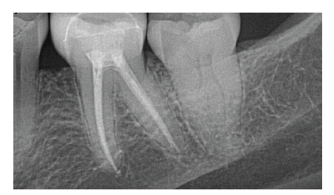
Non-surgical endodontic therapy can be achieved by various filing and obturation methods, however certain core principles must be adhered to. The canal system must be debrided of bacteria and contaminants, it must be disinfected, and the root apex must be appropriately sealed along with the coronal aspect of the tooth(1). In order to perform this therapy successfully, the clinician must have a proper foundation in each of these principles and must also comprehend tooth morphology and associated varying complexities(2,3,4,5,6). Weine, Vertucci, and Ahmed, among others, established classification systems for root canal morphology that denote the commonly occurring canal configurations(4,5,6) (Table 1), but the challenge still remains in how to best manage and treat these complex anatomical variances. Several factors such as the number of roots, the number of canals, canal classification, presence of stenosis and or calcifications, sharp curvatures, lateral canals, and atypical apices, can affect the success rate of non-surgical endodontic therapy. And while even though the use of proven clinical techniques and sealers can still result in failed endodontic therapy(1), advancements in techniques and materials are helping to mitigate this incidence of failure. CBCT imaging has also played a major role in the advancement of endodontics by allowing clinicians to more effectively diagnose cases and identify and recognize tooth and canal morphology and anatomy.
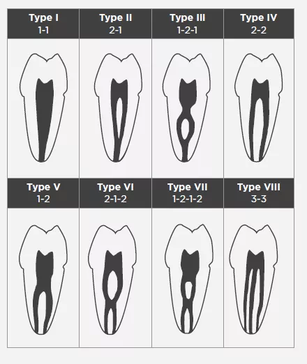
Table 1. Vertucci’s Canal Classification
Type I: a single canal extends from the pulp chamber to the apex.
Type II: two separate canals leave the pulp chamber and join short of the apex to form one canal.
Type III: one canal leaves the pulp chamber, divides into two within the root, and then merges to exit as one canal.
Type IV: Two separate and distinct canals extend from the pulp chamber to the apex.
Type V: one canal leaves the pulp chamber and divides short of the apex into two separate and distinct canals with separate apical foramina.
Type VI: two separate canals leave the pulp chamber, merge in the body of the root, and re-divide short of the apex to exit as two distinct canals.
Type VII: one canal leaves the pulp chamber, divides and then rejoins within the body of the root, and finally re-divides into two distinct canals short of the apex.
Type VIII: three separate and distinct canals extend from the pulp chamber to the apex.
For the past 30 years, a combination of hand files and nickel titanium rotary files have been used to instrument and debride the canals to then allow for delivery of disinfecting irrigants into the canal system and subsequent obturation. A widely accepted technique is to pre-enlarge the coronal and middle thirds of the root to then perform scouting and final instrumentation of the apical third. This technique allows for adequate debris removal, introduction of irrigants, with less risk of the file binding in multiple points which can lead to separation and or potential for intra-canal fractures(7). And while the metallurgy of these NiTi rotary files allow them to be stronger and more flexible than their stainless steel counterparts, they can still be susceptible to breakage, even without prior use or signs of deformation(8.9).
Utilization of nickel titanium files with this technique is still considered to be advantageous over stainless steel; however, some of the risks and disadvantages to using them in this method include: over instrumentation and removal of dentin leading to thinning of the root canal walls and potential for subsequent fracture, straightening and transportation of canal and apex, ledging within the canal, extrusion of debris, apical blockage, and file separation(10).
Current concepts of endodontic therapy place an emphasis on glide path, conservative dentin removal and flexibility in their file designs in an attempt to preserve as much as the natural tooth anatomy as possible. Glide path can reduce these aforementioned risks by facilitating canal shaping during the instrumentation process while following the existing path of the canal(10,11). Canal anatomy and curvatures vary greatly, and curvatures greater than 30°(12,10) are considered significant and can pose significant challenges and risks such as file separation, canal straightening and transportation, ledging, and stripping and perforation. Because of this, glide path management can be a crucial step in the outcome of endodontic therapy.
Many manufactures have developed thermally treated file systems to conservatively perform endodontic therapy by designing efficiently cutting, safe-ended, flexible files with an offset center of rotation, controlled memory, and a maximum flute diameter that allow the files to follow the natural anatomy and curvature of the canal, while at the same time these improvements reducing the risk of file separation due to cyclic fatigue and torsional stress. The goal of these modifications is to improve the overall success of endodontic therapy, provide efficient cutting, preserve the natural shape of the canal, and resist fracture of the file even in the most demanding of circumstances of narrow and curved root canals. Sufficiently instrumented canals allow irrigants to be introduced and activated for three dimensional disinfection and subsequent obturation(13).
A comparative review of three file systems, each having different characteristics and approaches for treating complex anatomy, was performed. The file systems evaluated were Dentsply’s ProTaper Gold, ProTaper Next, and Kerr’s ZenFlex.
Thirty-six extracted molars with roots having curvatures between 30°-60° and similar anatomy were randomly selected. Twelve root canals were performed with each file system following each manufacturer’s settings and instructions for use, and a new set of files were used on each tooth respectively. Straight line access was established, and each tooth was initially instrumented with hand files (Kerr K-flex) up to a size 10 to working length using Kerr’s Elements E-motion motor with handpiece ratio of 8:1. Kerr’s Traverse orifice opener and glide path files, and Dentsply’s SX orifice opener and ProGlider glide path files were each used in conjunction with their associated rotary file systems. Copious irrigation performed throughout and in between each file with a side-vented syringe canula using sterile saline, and Kerr SlickGel ES was used as lubricant. Recapitulation was performed with a size 10 K-file to maintain patency. Rotary files were cleaned of debris and SlickGel reapplied between insertion into each canal. Rotary files were used in a brushstroke fashion, and files were already rotating when iinserted into the tooth.
Master apical files size varied and was dependent upon the canal and tooth and ranged from 20- 30. One of the reasons for this was because the Dentsply file systems ranged in size but did not exceed size 30 in their assorted packs. ProTaper Next went up to a X3 [30/0.07] in its assorted pack and ProTaper Gold went up to a F3 [30/0.09] in its assorted pack. Larger sizes of the Dentsply files are offered but not included in the assorted pack. The Zenflex assorted file packs go from 20/0.04 to a size 45/0.04 and to 45/0.06 respectively; however, only the 0.04 taper files were utilized in this comparison. Kerr Tubli-Seal Xpress was used for sealer along with each manufacturer’s respectively matching fitted gutta-percha for obturation.
Each file system was carefully assessed as to how it handled complex anatomy to reach working length, and digital radiographs were taken to assess their effectiveness to reach working length and their impact on the final shape of the canals.
Dentsply Pro-Taper Gold®
Dentsply ProTaper Gold files work by utilizing a progressive taper to sequentially preflare the coronal and middle thirds of the canal with shaping files and then separately instrument the apical third with finishing files in a stepwise fashion(13,14,15). This allows for the apical third to be instrumented without interference or engagement of the canal walls in the upper two thirds, thereby reducing the torsional stress and cyclic fatigue on the file(13,14,15). The files have non-end cutting tips, convex, triangular cross-sections with a changing helical angle and pitch over their cutting blades, and rotate on their center of mass(14). The Gold-wire technology in the ProTaper Gold files is a post-machining heat treatment process that allows for increased flexibility and strength and makes them more resistant to fracture when compared to ProTaper Universal(13).
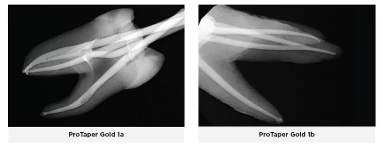
During this trial, it was observed that the downside to this technique is that it can widen the canal too much and subsequently weaken the canal walls which may lead to root fracture, canal transportation, zipping, and perforation. This is especially true in canals with multiple multi-plane curvatures. Moreover, the flexibility decreases and file memory increases as you go up in file size above a 25 file; therefore, ledging, transporting, and straightening of the canal was noted. Because the file rotates on its own center of mass, it constantly engages the canal walls, which often results in the file being sucked in (taper lock) and can lead to binding and fracture of the file. This also lends itself to a lot of rotational force being applied to the tooth and files, especially the finishing files as they increased in size.
It was also observed that when the ProGlider glide path file was used, it made it easier to negotiate the canals and instrument them to working length and reduced the load being placed on the initial ProTaper Gold shaping files. When scouting tight and constricted canals, the Gold SX orifice opener would unwind and strip easily, as the tip is very thin and fine. This file deformation is intended to prevent the file from breaking, however if unnoticed, the file will separate.
When the canals were obturated to the corresponding Pro-Taper Gold files, the matching gutta-percha typically fit well and went to working length, with the exception of three canals where the gutta-percha extruded well out of the apex, even in situations where the file did not nor could not pass out the apex. In those situations, the gutta-percha was trimmed to fit the working length. Radiographic examination of the teeth revealed three other canals where the gutta-percha was short approximately 0.5-1mm.
ProTaper Next®
Similar to the ProTaper Gold design, the ProTaper Next files have a progressively percent tapered design on a single file(16). However, the ProTaper Next files use M-wire technology which improves resistance to cyclic fatigue, decreases the risk for file separation, and increases flexibility(16). Additionally, these files have an offset mass of rotation so that the file contacts the canal wall at 2 points(16). This offset mass of rotation decreases taper lock, allows more cross-sectional space for more efficient cutting and debris removal, and allows a smaller more flexible file to cut a same sized preparation as a larger and stiffer file with a centered mass and axis of rotation(16).
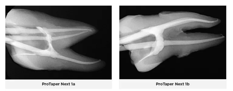
ProTaper Next files with M-Wire technology have been shown to cut dentin very well, but with subsequent use, there might be significant coronal, mid-root and apical canal transportation as well as an increase in the overall canal volume(17). This results in a loss of the original anatomy of the canal.
Observations noted when using ProTaper Next during this trial were that the file system cuts more efficiently and is more efficient in the number of steps than the ProTaper Gold system. The X1 and X2 files were more flexible than the X3, however, without controlled memory, they would still straighten out and transport the canal. The X3 file grabs and binds the tooth structure even more and takes away from the natural contour and anatomy. The ProGlider file followed the natural anatomy of the canal much more so than the ProTaper Next files. Because they lack controlled memory, the files are more likely to ledge and transport the canal, and they also tend to bind along multiple points within the canal which leads to an increased risk of separation. They do a lot of chipping and cutting of dentin from the canal walls, so you constantly have to irrigate out debris and recapitulate; because if not, you can easily lose patency.
Obturation with the fitted gutta-percha went to working length and fit the canals well in most cases; however, the gutta-percha extended out the apex in one canal because of a larger anatomical foramen, and the gutta-percha stopped short in six other canals. Three of those canals had ledging caused by the X1 and X2 files and patency could not be regained, so the gutta-percha could not reach the full working length. And, in one instance, the gutta-percha stopped short, but upon re-instrumentation of the canal three more times, the gutta-percha was able to be seated to the full working length.
ZenFlex™
ZenFlex files are nickel-titanium files that have undergone a patented heat treatment to provide them with a blend of flexibility and strength not typically seen in a file with this small mass. These files are designed to be as resistant or more resistant to torsional stresses relative to major rotary file competitors despite having a smaller maximum flute diameter and smaller mass. Two unique features that differentiate ZenFlex files from ProTaper Gold and ProTaper Next files are their controlled memory and maximum flute diameter of 1mm. ZenFlex files undergo a proprietary heat treatment process that improves their strength and resistance to cyclic fatigue, torsional stress and separation, as well as making them significantly more flexible and able to retain their cutting efficiency by preserving their sharp cutting edge. Unlike other manufacturers, Kerr uses a proprietary double heat treatment process. The files are heat treated first, their shape is then ground into them, and then they undergo a second heat treatment to make them stronger and sharper. They also heat treat each file differently based on its mass, therefore the optimum qualities for each file are obtained: smaller more flexible files become stronger, and larger stronger files become more flexible. The files come in both 0.04 and 0.06 tapers, have a non-end cutting tip, and are extremely strong and flexible with controlled memory. Their controlled memory and 1mm maximum flute diameter enable them to follow the natural contour of the canal and be minimally invasive to successfully instrument the canal without removing too much dentin from the coronal and middle thirds and thus preserve the integrity of the tooth structure. ZenFlex files have a centered mass of rotation with a triangular cross-section to provide excellent cutting and debris removal with little to no apical debris extrusion.
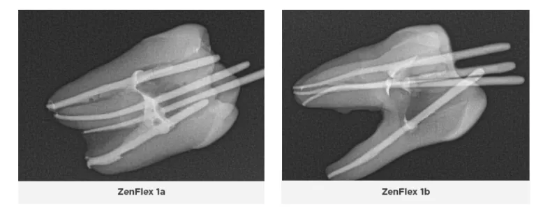
It was immediately observed that the 0.04 tapered ZenFlex files used in this comparison trial cut much more efficiently and allow the clinician to treat the tooth in a more dynamic and flexible workflow if one chooses to do so. Therefore, the clinician can use his or her preferred workflow and technique to instrument the canal.
Moreover, the time to treat each tooth was 30-50% faster than the ProTaper Gold and Next and file systems because the ZenFlex files cut more efficiently. Noticeably less binding and rotational force was applied to the tooth when using the ZenFlex files. Hardly any apical extrusion of debris was noted, and debris was observed to be removed coronally. The natural anatomy of the canals was preserved, and no evidence of transportation, ledging, nor blocking of any of the canals was observed.
In situations when the canals are constricted and stenotic, a lot of filing must typically be performed to establish a glide-path to working length before a rotary file can be taken to length(10,11,12,10). However, the ZenFlex files cut so well that, along with their flexibility and controlled memory, clinicians can negotiate these types of scenarios quickly using ZenFlex files in conjunction with the Traverse Orifice Opener and Glide Path files, difficult cases take much less time and can be performed more efficiently without destroying the anatomy of the tooth or separating files.
The Medium Fine (MF) 0.04 taper Kerr AutoFit Greater Taper gutta-percha was used with the 0.04 taper ZenFlex files, and had to be trimmed to the corresponding master apical file size. Once fitted and trimmed, there were no issues seating the gutta-percha to the established working length. Should a clinician use the 0.06 taper ZenFlex files, the AutoFit Greater Taper gutta-percha is also available in a 0.06 taper to match.
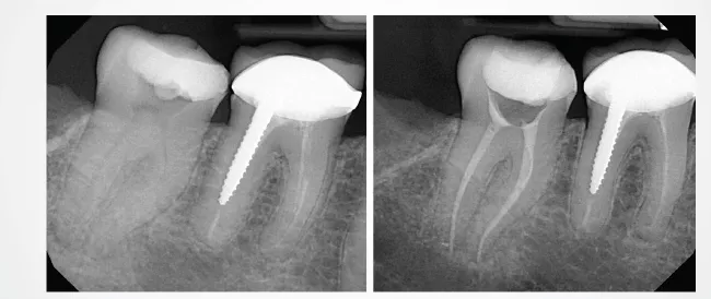
Two dimensional radiographs were taken of the teeth after endodontic therapy was completed and reviewed. In some of the ProTaper Gold and Next cases, working length was not reached radiographically, even though it was thought to be achieved clinically. As an electronic apex locator cannot be used to assess working length on extracted teeth, working length was established with visual observation. Clinically, an electronic apex locator would have been used, and adjustments would have been made throughout the entire process to ensure the working length was reached. The teeth were selected at random, and some of the orifices and canals were calcified or stenotic. In a live clinical scenario, ultrasonics would have been used to negotiate these constrictions more easily; however that being said, the ZenFlex and Traverse file systems allowed these canals to be negotiated and treated without issue. Additionally, the teeth treated with the ZenFlex files appeared to have the best preservation of the natural shape of the canal and conserved the most tooth structure. This was not the case with the ProTaper Gold and Next file systems. Less efficient cutting with more aggressive removal of tooth structure was noted, resulting in a loss in the original anatomy of the canals. ZenFlex is an ideal file that balances strength and flexibility to allow clinicians to reach the working length while being minimally invasive, even in cases with complex canal anatomies.
Because human teeth exhibit a high degree of variability in canal anatomy compared to artificially simulated canals of resin blocks(18), best efforts were made to achieve similar anatomical consistency among the three groups.
Human teeth are preferred over resin blocks as extracted human teeth have similar micro-hardness to natural dentin(19). Moreover, the heat generated from the files could soften the resin block matrix causing the files to bind, thereby altering the outcome (19,20). Due to these limitations in the study, further research and clinical evaluation is needed to evaluate the efficacy of the files. CBCT and or SEM analysis of the canal shapes, transportation, integrity of the tooth structure, as well as file integrity and deformation would further examine the properties of these files.
Conclusion
I am highly impressed by Kerr’s ZenFlex file system’s ability to endodontically treat all cases, from simple to complex, using the clinician’s preferred technique. I especially love how it perseveres the integrity of the tooth structure and natural anatomy with its controlled memory and 1mm maximum flute diameter. The proprietary variable heat treatment based on mass makes these files incredibly strong and flexible, provides exceptional cutting efficiency, and significantly reduces the risk for separation. With Kerr ZenFlex files, clinicians can provide optimal, minimally invasive endodontic therapy.
Disclosure: Dr. Miller has received an honorarium for his participation in this study.
ZenFlex is a trademark of Kerr Corporation. All other trademarks are properties of their respective owners.
The opinions and techniques discussed are based on the experience of Dr. Matthew Miller. Kerr is a medical device manufacturer
and does not dispense medical advice. Clinicians should use their own judgment in treating their patients.
Bibliography
1. Christensen, Gordon J. Clinicians Report. Volume 10 Issue 3, March 2017, Pages 1-3.
2. Pamukcu Guven, Esra. Root Canal Morphology and Anatomy. July 7, 2019. doi:10.5772/intechopen.86096.
https://www.intechopen.com/chapters/67419
3. Cleghorn BM, Christie WH, Dong CC. Anomalous mandibular premolars: a mandibular first premolar with three roots and
a mandibular second premolar with a C-shaped canal system. Int Endod J. 2008 Nov;41(11):1005-14. doi: 10.1111/j.1365-
2591.2008.01451.x. PMID: 19133090.
4. Weine FS, Healey HJ, Gerstein H, Evanson L. Canal configuration in the mesiobuccal root of the maxillary first molar and its
endodontic significance. Oral Surg Oral Med Oral Pathol. 1969 Sep;28(3):419-25. doi: 10.1016/0030-4220(69)90237-0. PMID:
5257186.
5. Vertucci FJ. Root canal anatomy of the human permanent teeth. Oral Surg Oral Med Oral Pathol. 1984 Nov;58(5):589-99. doi:
10.1016/0030-4220(84)90085-9. PMID: 6595621.
6. Ahmed HMA, Versiani MA, De-Deus G, Dummer PMH. A new system for classifying root and root canal morphology. Int Endod J.
2017 Aug;50(8):761-770. doi: 10.1111/iej.12685. Epub 2016 Oct 17. Erratum in: Int Endod J. 2018 Oct;51(10):1184. PMID: 27578418.
7. Ruddle, Clifford J. Current concepts for preparing the root canal system. Dentistry Today. 2001 Feb;20(2):76-83. PMID: 12524850.
8. Pruett JP, Clement DJ, Carnes DL Jr. Cyclic fatigue testing of nickel-titanium endodontic instruments. J Endod. 1997
Feb;23(2):77-85. doi: 10.1016/S0099-2399(97)80250-6. PMID: 9220735.
9. Gambarini G. Cyclic fatigue of nickel-titanium rotary instruments after clinical use with low- and high-torque endodontic
motors. J Endod. 2001 Dec;27(12):772-4. doi: 10.1097/00004770-200112000-00015. PMID: 11771588.
10. Cassim I, van der Vyver PJ. The importance of glide path preparation in endodontics: a consideration of instruments and
literature. SADJ. 2013 Aug;68(7):322, 324-7. PMID: 24133951.
11. Plotino G, Nagendrababu V, Bukiet F, Grande NM, Veettil SK, De-Deus G, Aly Ahmed HM. Influence of Negotiation, Glide Path,
and Preflaring Procedures on Root Canal Shaping-Terminology, Basic Concepts, and a Systematic Review. J Endod. 2020
Jun;46(6):707-729. doi: 10.1016/j.joen.2020.01.023. Epub 2020 Apr 22. PMID: 32334856.
12. Szep S, Gerhardt T, Leitzbach C, Lüder W, Heidemann D. Preparation of severely curved simulated root canals using engine-driven
rotary and conventional hand instruments. Clin Oral Investig. 2001 Mar;5(1):17-25. doi: 10.1007/pl00010680. PMID: 11355093.
13. Ruddle CJ, Machtou P, West JD. Endodontic canal preparation: new innovations in glide path management and shaping canals.
Dent Today. 2014 Jul;33(7):118-23. PMID: 25118526.
14. Ruddle, Clifford J. (2005). The ProTaper technique. Endodontic Topics. 10. 187 - 190. doi: 10.1111/j.1601-1546.2005.00115.x.
15. West J. Progressive taper technology: rationale and clinical technique for the new ProTaper universal system. Dent Today. 2006
Dec;25(12):64, 66-9. PMID: 17193790.
16. Ruddle, Clifford J. (2021). ProTaper Next System. Advanced Endodontics. https://www.endoruddle.com/ProTapernext.
17. van der Vyver PJ, Paleker F, Vorster M, de Wet FA. Root Canal Shaping Using Nickel Titanium, M-Wire, and Gold Wire: A Microcomputed
Tomographic Comparative Study of One Shape, ProTaper Next, and WaveOne Gold Instruments in Maxillary First
Molars. J Endod. 2019 Jan;45(1):62-67. doi: 10.1016/j.joen.2018.09.013. Epub 2018 Nov 13. PMID: 30446405.
18. Dummer PM, Alodeh MH, al-Omari MA. A method for the construction of simulated root canals in clear resin blocks. Int Endod
J. 1991;24:63–66.
19. Lim YJ, Park SJ, Kim HC, Min KS. Comparison of the centering ability of Wave·One and Reciproc nickel-titanium instruments in
simulated curved canals. Restor Dent Endod. 2013;38:21–25.
20. Yoo YS, Cho YB. A comparison of the shaping ability of reciprocating NiTi instruments in simulated curved canals. Restor Dent
Endod. 2012;37:220–227.
MKT-22-0114
Share This


